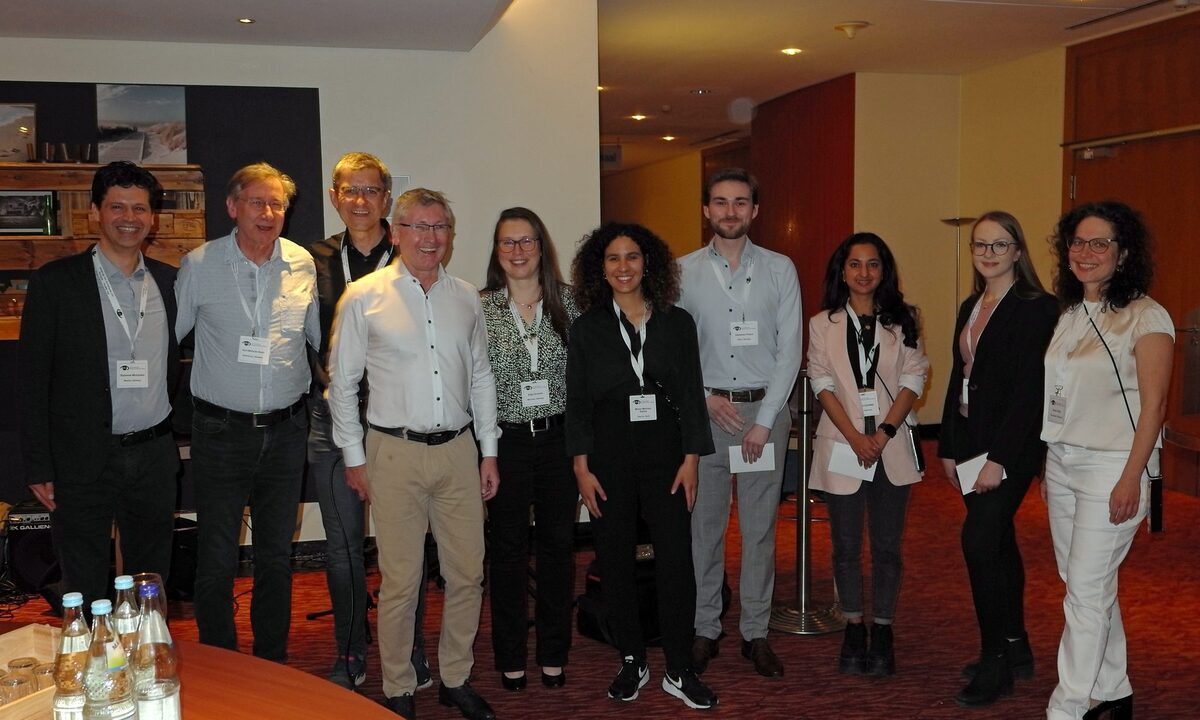
Posterpreisverleihung beim Potsdam Meeting 2024 der Stiftung zur Verhütung von Blindheit
So viele Forscherinnen und Forscher wie noch nie zuvor, haben die Möglichkeit genutzt ihre Arbeiten zur Forschung beim 18. Internationalen Forschungskolloquium zur Retinalen Degeneration in Potsdam einzureichen.

Das Internationales Forschungskolloquium zu Netzhauterkrankungen fand vom 12. bis 13. April 2024 in Potsdam statt. Eine sehr wichtige Funktion dieser Tagung ist die Posterpräsentation. Die Abstracts für die Posterpreise konnten dort vor einem internationalen Publikum präsentiert werden. Sie zeigten das Engagement für die Forschung und sicherten sich damit die Chance auf renommierte Auszeichnungen.
Im Namen der Pro Retina – Stiftung zur Verhütung von Blindheit bedankt sich das Organisationskomitee und die Jury für die vielen hervorragenden Beiträge und gratuliert den Preisträgern.
Hier die ausgezeichneten Poster:
Role of exosomal miR-205-5p in angiogenesis and migration
Miriam Martínez-Santos 1,3, María Ybarra 1,3, María Oltra2,3, Maria E. Pires1, Chiara Ceresoni1, Jorge M. Barcia1,2,3* , Javier Sancho-Pelluz2,3
1 Escuela de Doctorado, Universidad Católica de Valencia, 46001 Valencia, Spain / 2 Facultad de Medicina y Ciencias de la Salud, Universidad Católica de Valencia, 46001 Valencia, Spain / 3 Centro de Investigación Traslacional San Alberto Magno, Universidad Católica de Valencia, 46001 Valencia, Spain
Purpose:
Small extracellular vesicles (sEVs) represent a pivotal component in intercellular communication, carrying a diverse array of biomolecules. The dynamics of sEVs release are significantly influenced by the cellular microenvironment such as high glucose (HG). This phenomenon has been associated with the promotion of pathological processes. This study aims to understand the role of miR-205-5p in extracellular vesicles under hyperglycemic conditions and its effect on angiogenesis and cell migration.
Fluorescence lifetime imaging microscopy (FLIM) of pigmented cells in neovascular age-related macular degeneration (AMD)
Sebastian Fritsch, Katharina Wall, Leon von der Emde, Frank G. Holz, Christine A. Curcio, Thomas Ach
Department of Ophthalmology, University Hospital Bonn, Bonn, Germany / Department of Ophthalmology, University of Alabama at Birmingham, Birmingham, AL, USA
Purpose:
Melanotic cells are often found in fibrotic tissue in the outer retina in the course of late stage neovascular AMD. De novo synthesis of melanin or conversion of individual organelles in the context of epithelial-mesenchymal-transition (EMT) of the retinal pigment epithelium (RPE) are possible mechanisms in the formation of these pigmented lesions, but the pathogenesis and impact of these lesions is not yet fully understood. Fluorescence lifetime imaging microscopy (FLIM) enables to examine underlying fluorophores or groups of fluorophores in autofluorescent tissue. This study uses FLIM to examine these pigmented lesions and to further determinethe origin of the pigment.
Vascularization of human stem cell-derived 3D retinal organoids
Kritika Sharma 1, Bohao Cui 2, Carmen Ruiz de Almodóvar 2, Volker Busskamp 1,
1Augenklinik-Bonn, Ernst-Abbe-Str. 2, 53127-Bonn. / 2Life and Brain GmbH, Venusberg-Campus 1, 53127 Bonn.
Purpose:
Retinal organoids (ROs) are 3D in vitro structures that attempt to recapitulate the development, structure, and functioning of in vivo human retina. RO field has evolved over time meticulously regulating features of organoid protocol like structural complexity, scalability and reproducibility. However, inefficient exchange of oxygen and nutrients and periodic cellular waste removal generates an expanding necrotic core causing aggressive ganglion cell (GC) death resulting in premature functional decline. Vasculature within the developing organoid has shown to combat above mentioned drawbacks. Here, we make use of an efficient hiPSC-derived inducible endothelial cell (EC) differentiation protocol that gives rise to an endogenous, self-organizing vascular-like system within the retinal layers prolonging the survival and long-term functioning of RO.
Inflammatory protein profiling in aqueous humour of patients with non-exudative age-related Macular Degeneration
Juliane Schikora1, Aaron Dort 1, Hannah N. Wolf 1, Mihály Józsi 2, Richard Pouw 3, Thomas Bertelmann 4, Dirk Bahlmann 4, Christian van Oterendorp 4, Nicolas Feltgen 5, Hans Hoerauf 4, Jannis Klemming 4, Diana Pauly 1
1 Experimental Ophthalmology, University Marburg, Marburg, Germany / 2 Immunology, ELTE Eötvös Loránd University, Budapest, Hungary / 3 Sanquin Research, Amsterdam, Netherlands / 4 Ophthalmology, University Medical Centre Göttingen, Göttingen, Germany / 5 Ophthalmology, University Basel, Basel, Switzerland
Purpose:
A dysregulated immune system, particularly the complement system, plays a fundamental role in the pathophysiology of age-related macular degeneration (AMD). The aim of this study was to analyse complement components and inflammatory mediators in the aqueous humour of non-exudative AMD patients and to compare them with a control group.
Die vollständige Beschreibung der eingereichten Abstracts finden Sie im Conference Report.
18th PRO RETINA Research-Colloquium Potsdam 2024
Retinal Degeneration – Advancing Retinal Research